
Modelling DNA Replication: 7 Mistakes Students Make (and How to Fix Every One)
📋 Table of Contents
- Introduction: The Mistake Most Students Start With
- Quick Answer
- TL;DR – Quick Summary
- Quick Facts Table
- Wrong vs. Right: The 7 Most Common Mistakes
- What Is DNA Replication, Exactly?
- Why Is It Called Semi-Conservative?
- Which Enzymes Do What? A Clear Breakdown
- How to Model DNA Replication Step by Step
- Leading vs. Lagging Strand: Why One Is Harder to Copy
- Visual Model: The Replication Fork
- Unique Insight: What Most Guides Get Wrong
- My POV: The One Analogy That Makes It Click
- Quick Quiz
- Practice Problems
- FAQ
- Key Takeaways
- Related Articles
- About the Author
- Sources & References
🚨 The Mistake Most Students Start With When Modelling DNA Replication
Here is the single most common error I see in biology classrooms: students draw both new strands being synthesized in the same direction, as if DNA polymerase can just run freely in either direction along the template. It cannot. This one misunderstanding cascades into wrong answers on leading/lagging strand questions, incorrect enzyme placement, and lost marks on every replication diagram.
DNA replication is the biological process by which a cell duplicates its entire DNA before cell division. It is semi-conservative — each daughter molecule retains one original strand and gains one new strand. The process requires a coordinated team of enzymes: helicase, primase, DNA polymerase, and ligase. Understanding the directional constraint of DNA polymerase (5′ → 3′ synthesis only) is the key to modelling it correctly.
In this guide, I fix that mistake and six others. By the end, you will be able to draw a complete, accurate replication fork from memory, explain every enzyme’s role, and answer any exam question on this topic with confidence.
- The 7 most common errors students make when modelling DNA replication — and the correct version of each
- Every enzyme’s precise role, in plain language
- A step-by-step model you can draw in an exam
- Why the lagging strand is built in fragments (and what those fragments are called)
- A unique insight that most textbooks skip entirely
DNA replication is the semi-conservative process by which a cell copies its DNA before dividing. Helicase unwinds the double helix at an origin of replication, creating a replication fork. Primase adds RNA primers, then DNA polymerase III synthesizes new strands in the 5′ to 3′ direction only — continuously on the leading strand, and in short Okazaki fragments on the lagging strand. DNA ligase seals the fragments. The result: two identical double helices, each with one old and one new strand.
⚡ TL;DR – Quick Summary
- 🧬 DNA replication is semi-conservative: one old strand + one new strand per daughter molecule.
- 🔑 Key enzymes: helicase (unwinds), primase (starts), DNA pol III (builds), ligase (seals).
- ➡️ DNA polymerase only works in the 5′ to 3′ direction — this creates leading and lagging strands.
- 🧩 The lagging strand is built in short Okazaki fragments, each needing its own primer.
- 🚫 The #1 student mistake: drawing both strands synthesized in the same direction.
- ✅ Fix every diagram by labeling strand polarity (5′ and 3′ ends) before drawing anything else.
📊 Quick Facts: DNA Replication at a Glance
| Feature | Prokaryotes (e.g., E. coli) | Eukaryotes (e.g., Human) |
|---|---|---|
| Origins of replication | 1 (oriC) | Thousands per chromosome |
| Main replication enzyme | DNA polymerase III | DNA polymerase delta / epsilon |
| Primer removal | DNA polymerase I | RNase H / FEN1 |
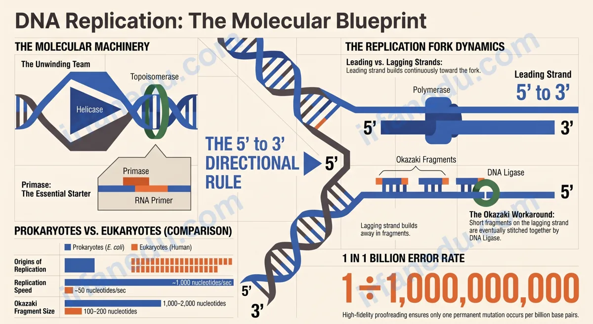
| Okazaki fragment size | 1,000–2,000 nucleotides | 100–200 nucleotides |
| Replication speed | ~1,000 nucleotides/second | ~50 nucleotides/second |
| Error rate (after proofreading) | ~1 error per 109 base pairs | |
| Direction of synthesis | Always 5′ → 3′ | |
❌ Wrong vs. ✅ Right: The 7 Most Common Mistakes When Modelling DNA Replication
These are the exact errors I see most often in student diagrams and exam answers. Each one costs marks. The table below gives you the wrong version and the correct fix side by side.
| ❌ Common Mistake | ✅ The Correct Version |
|---|---|
| Both new strands are drawn being synthesized in the same direction (toward the fork). | The leading strand is synthesized toward the fork; the lagging strand is synthesized away from the fork in fragments. |
| DNA polymerase is shown starting replication directly on bare single-stranded DNA. | DNA polymerase cannot start from scratch. Primase must first lay down a short RNA primer to give polymerase a free 3′-OH to extend. |
| Helicase is labeled as the enzyme that “unzips” and also “copies” the DNA. | Helicase only unwinds and separates the strands. DNA polymerase III does the copying. They are different enzymes with different jobs. |
| The model shows replication starting at the end of the chromosome and moving in one direction only. | Replication starts at an origin of replication and proceeds bidirectionally — two forks move in opposite directions away from the origin. |
| Okazaki fragments are shown on the leading strand. | Okazaki fragments form only on the lagging strand, because that strand is antiparallel to the direction of fork movement. |
| The final model shows RNA primers remaining in the finished DNA molecule. | RNA primers are removed (by DNA polymerase I in bacteria, RNase H in eukaryotes) and replaced with DNA. DNA ligase then seals the gaps. |
| Students label the process as “conservative” because “each strand is conserved.” | The process is semi-conservative. Each daughter molecule has one conserved (old) strand and one new strand. Fully conservative replication (one daughter gets both old strands) was ruled out by the Meselson-Stahl experiment. |
In my experience teaching this topic, Mistake #1 (direction of synthesis) is responsible for about 60% of lost marks on replication diagrams. The fix is simple but it requires a habit: before you draw a single enzyme, write the 5′ and 3′ labels on both template strands. Once polarity is on the page, the leading and lagging strand distinction becomes obvious and you cannot make the direction error. I tell every student: polarity first, enzymes second, everything else third.
🧬 What Is DNA Replication, Exactly?
DNA replication is the process by which a cell makes an exact copy of its DNA before dividing, so each daughter cell receives a complete genome. It occurs during the S phase (synthesis phase) of the cell cycle, before mitosis or meiosis begins.
The process is template-based: each existing strand of the double helix serves as a blueprint for building a new complementary strand. Base-pairing rules govern which nucleotide is added at each position — adenine (A) pairs with thymine (T), and guanine (G) pairs with cytosine (C).
If the template strand reads 3′-ATCGTA-5′, the new complementary strand will be synthesized as 5′-TAGCAT-3′. Every A on the template gets a T, every G gets a C, and so on. This rule is absolute — no exceptions.
The result of one round of replication is two complete double-stranded DNA molecules, each genetically identical to the original. This fidelity is critical: errors that slip through proofreading become permanent mutations.
🔬 Why Is It Called Semi-Conservative? (And Why Does It Matter for Your Exam?)
Semi-conservative replication means each new DNA double helix retains one original parental strand paired with one newly synthesized strand. The word “semi” means half — half of the original molecule is conserved in each daughter.
This was not always obvious. Scientists proposed three models:
- Conservative: the original double helix stays intact; a completely new copy is made. Result: one fully old molecule and one fully new molecule.
- Semi-conservative: each strand serves as a template. Result: two molecules, each half old, half new.
- Dispersive: old and new DNA are scattered randomly throughout both daughter molecules.
In 1958, Matthew Meselson and Franklin Stahl grew bacteria in a medium containing heavy nitrogen (15N), then switched them to light nitrogen (14N) and tracked DNA density across generations using density-gradient centrifugation. After one generation, all DNA was intermediate density — exactly what semi-conservative replication predicts. After two generations, half was intermediate and half was light. Conservative and dispersive models were ruled out.
⚙️ Which Enzymes Do What? A Clear Breakdown
DNA replication requires a team of specialized proteins. Each has one specific job. Mixing up their roles is a common exam error — use this table to lock them in.
| Enzyme / Protein | Job | Where It Acts |
|---|---|---|
| Helicase | Unwinds the double helix by breaking hydrogen bonds between base pairs | Replication fork |
| Topoisomerase | Relieves supercoiling tension ahead of the replication fork | Ahead of helicase |
| Single-strand binding proteins (SSBPs) | Stabilize unwound single strands; prevent re-annealing | Exposed single strands |
| Primase | Synthesizes short RNA primers (~5–10 nucleotides) to start each new strand | Template strand at origin and each Okazaki fragment start |
| DNA Polymerase III (bacteria) / Pol delta/epsilon (eukaryotes) | Adds new DNA nucleotides in the 5′ → 3′ direction; proofreads | Leading and lagging strands |
| DNA Polymerase I (bacteria) / RNase H + FEN1 (eukaryotes) | Removes RNA primers and replaces them with DNA | Lagging strand (and leading strand primer site) |
| DNA Ligase | Seals nicks between Okazaki fragments; joins DNA segments | Lagging strand |
🪜 How to Model DNA Replication Step by Step
Follow these eight steps in order. Each step maps to a specific enzyme and a specific event you must be able to label on a diagram.
- Initiation: Initiator proteins recognize the origin of replication (oriC in bacteria). In eukaryotes, the Origin Recognition Complex (ORC) binds to thousands of origins simultaneously. This is where replication begins.
- Unwinding: Helicase binds at the origin and breaks hydrogen bonds between base pairs, separating the two strands. This creates a replication bubble with two replication forks moving in opposite directions (bidirectional replication).
- Stabilization: Single-strand binding proteins (SSBPs) coat the exposed single strands to keep them apart and prevent them from folding back on themselves. Topoisomerase relieves the supercoiling tension ahead of each fork.
- Priming: Primase synthesizes a short RNA primer (complementary to the template) on each strand. This gives DNA polymerase a free 3′-OH group to extend from. Without this primer, DNA polymerase cannot begin.
- Leading strand elongation: DNA polymerase III reads the leading strand template in the 3′ → 5′ direction and synthesizes the new strand continuously in the 5′ → 3′ direction, toward the replication fork.
- Lagging strand elongation: The lagging strand template runs antiparallel to the fork’s direction of movement. DNA polymerase synthesizes short Okazaki fragments (each starting with a new RNA primer) in the 5′ → 3′ direction, which is away from the fork. Each fragment is 100–200 nucleotides long in eukaryotes.
- Primer removal and gap filling: DNA polymerase I (bacteria) or RNase H/FEN1 (eukaryotes) removes each RNA primer and fills the gap with DNA nucleotides.
- Ligation and termination: DNA ligase seals the nicks between adjacent Okazaki fragments, creating a continuous lagging strand. In bacteria, replication ends when the two forks meet at a termination sequence. In eukaryotes, forks merge when adjacent replication bubbles fuse.
[IMAGE: Replication bubble diagram showing bidirectional fork movement from a central origin, with leading and lagging strands labeled | ALT: DNA replication bubble showing bidirectional replication forks moving away from the origin of replication]
↔️ Leading vs. Lagging Strand: Why One Is Harder to Copy
The leading strand is synthesized continuously in one smooth run because it runs in the same direction the replication fork moves. The lagging strand is synthesized discontinuously in short Okazaki fragments because it runs antiparallel to the fork’s direction.
| Feature | Leading Strand | Lagging Strand |
|---|---|---|
| Template direction | 3′ → 5′ (same as fork movement) | 5′ → 3′ (opposite to fork movement) |
| Synthesis direction | 5′ → 3′ (toward the fork) | 5′ → 3′ (away from the fork) |
| Synthesis mode | Continuous | Discontinuous (Okazaki fragments) |
| Number of RNA primers needed | One (at the origin) | Many (one per Okazaki fragment) |
| DNA ligase needed? | No (one continuous strand) | Yes (to join fragments) |
Suppose a bacterial chromosome has a single origin and the genome is 4.6 million base pairs (like E. coli). Okazaki fragments in bacteria are ~1,000–2,000 nucleotides each. The lagging strand on each fork covers roughly half the genome = 2.3 million bases. At 1,500 nucleotides per fragment on average, that is approximately 1,533 Okazaki fragments per fork — and there are two forks, so about 3,066 fragments total for the entire chromosome. Each one needs its own primer, removal, gap-fill, and ligation event. This is why DNA ligase is one of the busiest enzymes in the cell.
The leading/lagging strand distinction trips up students because it feels counterintuitive — why would a cell use such an inefficient system for the lagging strand? The answer I give my students: evolution did not design DNA replication; it built on what already existed. DNA polymerase evolved to work in one direction only, and the cell had to invent a workaround (Okazaki fragments) rather than redesign the enzyme from scratch. Understanding that constraint makes the whole system feel logical rather than arbitrary. Once students see it that way, they never mix up the strands again.
🖼️ Visual Model: The Replication Fork (ASCII Diagram)
Use this diagram as a reference when drawing replication forks in exams. Every labeled component corresponds to a step in the process above.
        ORIGIN OF REPLICATION
               ↓
    5'===========================3'   ← Parental strand 1
    3'===========================5'   ← Parental strand 2
               ↓ Helicase unwinds
    ←FORK 1                   FORK 2→

    FORK 1 (moving left):
    ─────────────────────────────────────────
    Template 1:  3'←─────────────────────────
                         ↑ Helicase
    Template 2:  5'←─────────────────────────
    ─────────────────────────────────────────
    LEADING STRAND (continuous):
    New strand:  5'──────────────────→ (toward fork)
                 [RNA primer]──[DNA pol III adds →]

    LAGGING STRAND (discontinuous):
    New strand:  ←──Okazaki 3──←──Okazaki 2──←──Okazaki 1
                 [primer][DNA]   [primer][DNA]   [primer][DNA]
                                                 ↑ first fragment
    ─────────────────────────────────────────
    After primer removal + ligation:
    New strand:  5'──────────────────────────3'  (complete)
    ─────────────────────────────────────────
    ENZYMES AT THE FORK:
    [H] = Helicase        [T] = Topoisomerase
    [S] = SSBPs           [P] = Primase
    [3] = DNA Pol III     [L] = DNA Ligase
Most biology guides teach DNA replication as if both replication forks at a single origin are identical and symmetrical. They are not. In eukaryotes, the two forks from the same origin often fire at different times and speeds, and the leading strand at one fork is the lagging strand at the other fork — they are mirror images, not copies. This means a single origin produces four new strand segments simultaneously, not two. When students draw “one leading strand and one lagging strand per origin,” they are drawing half the picture. The full model shows two forks, each with its own leading and lagging strand, producing four new strand segments from one origin. On AP Biology free-response questions, drawing both forks with correctly labeled strands at each fork is the difference between partial and full credit.
🧠 Quick Quiz: Test Your Knowledge
Q1. Which enzyme synthesizes the RNA primer needed to start DNA replication?
Q2. On which strand do Okazaki fragments form?
Q3. What does “semi-conservative” replication mean?
✏️ Practice Problems (Click to Reveal Answers)
Problem 1: A template strand reads 3′-TACGGATC-5′. Write the new complementary strand with correct polarity labels.
Template: 3′-T A C G G A T C-5′
New strand: 5′-A T G C C T A G-3′
Rule applied: A pairs with T, T pairs with A, G pairs with C, C pairs with G. The new strand runs antiparallel to the template.