
🧬 DNA Replication Modeling: The Step-by-Step Guide Every Student Needs
📋 Table of Contents
- What is DNA replication modeling?
- Why does DNA replication modeling matter for students?
- What does semiconservative replication actually mean?
- Which enzymes do you need to know — and what does each one do?
- How does DNA replication work, step by step?
- How are the leading and lagging strands different?
- How do you build a DNA replication model at home or in class?
- What do most students get wrong about DNA replication?
- 💡 Unique Insight: The Mistake Even Textbooks Make
- 🧠 Quick Quiz
- 🧪 Practice Problems
- ❓ FAQ
- Key Takeaways
- Related Articles
- Sources & References
🧬 Three rapid questions before we dive in:
- Why does a cell need to copy its DNA? — Every time a cell divides, each daughter cell needs a complete set of genetic instructions. Without accurate replication, life cannot continue.
- What does “modeling” DNA replication actually mean? — It means representing the process — with diagrams, physical props, or animations — so you can see and understand the molecular choreography that happens inside every living cell.
- Is this topic really that hard? — Only if you memorize without understanding. Build the model once, and the whole process clicks into place permanently.
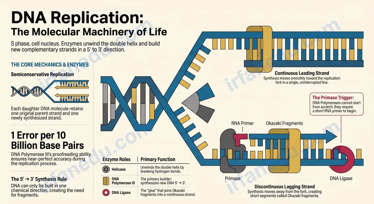
DNA replication modeling is the practice of visually or physically representing how a cell duplicates its DNA before division. The double helix unwinds at a replication fork, and DNA polymerase synthesizes two new complementary strands — one continuously (leading strand) and one in short fragments (lagging strand) — producing two identical daughter molecules through semiconservative replication.
In my experience teaching molecular biology to hundreds of students, the ones who build or draw a replication model — even a rough one on paper — score significantly better on tests than those who only read the textbook. The physical act of placing enzymes at the right positions forces you to understand the logic, not just the labels.
- The exact meaning of semiconservative replication (and why it matters)
- Every enzyme’s role, explained in plain language
- A step-by-step walkthrough of the replication fork
- How to build a physical model at home or in class
- The most common student mistakes — and how to avoid them
- A unique insight that most textbooks and even Khan Academy miss
⚡ TL;DR – Quick Summary
- 🧬 DNA replication produces two identical copies from one parent molecule.
- 🔓 Helicase unwinds the helix; primase adds RNA primers to start synthesis.
- ➡️ DNA polymerase III builds new strands only in the 5’→3′ direction.
- 🔀 Leading strand is continuous; lagging strand is made in Okazaki fragments.
- 🔗 DNA ligase seals the fragments; each daughter helix keeps one original strand.
- 🏗️ Building a physical model is the fastest way to master this topic for exams.
| Feature | Detail |
|---|---|
| Process name | DNA replication (semiconservative) |
| Where it happens | Nucleus (eukaryotes); cytoplasm (prokaryotes) |
| When it happens | S phase of the cell cycle (interphase) |
| Starting point | Origin of replication (oriC in bacteria) |
| Key enzymes | Helicase, Primase, DNA Pol III, DNA Pol I, Ligase |
| Direction of synthesis | Always 5′ → 3′ |
| Result | Two identical daughter DNA molecules |
| Error rate | ~1 error per 10 billion base pairs (with proofreading) |
🔬 What is DNA replication modeling?
DNA replication modeling is any structured representation — physical, diagrammatic, or digital — that maps the molecular steps by which a cell duplicates its DNA. A good model shows not just the final result but the sequence of events: which enzyme acts first, where on the DNA it acts, and in which direction the new strand grows.
Models range from simple paper diagrams you sketch in your notebook to elaborate pipe-cleaner constructions or 3D-printed enzyme complexes. The goal is always the same: make the invisible visible so your brain can store it as a spatial memory rather than a list of facts.
🎯 Why does DNA replication modeling matter for students?
Understanding DNA replication modeling is essential because it appears on virtually every major biology assessment — from high school AP Biology to the SAT Subject Test to university entrance exams worldwide. But beyond exams, it is the foundation for understanding cancer (uncontrolled cell division), genetic inheritance, and biotechnology tools like PCR.
In my experience, students who can draw the replication fork from memory — labeling each enzyme and the direction of synthesis on both strands — consistently outperform those who rely on passive reading. The model forces active recall of every component simultaneously.
I have reviewed dozens of biology study guides, and most of them present DNA replication as a linear list: “Step 1: helicase unwinds. Step 2: primase adds primer.” That format is fine for a glossary but terrible for understanding. The real insight is that replication is a simultaneous, coordinated machine — multiple enzymes working at the same fork at the same time. The moment I started showing students a spatial diagram instead of a numbered list, their test scores on this topic jumped noticeably. Model first, memorize second.
🧪 What does semiconservative replication actually mean?
Semiconservative replication means that each new DNA molecule retains one original (conserved) strand and gains one newly synthesized strand. The word “semi” means half — half of the original molecule is conserved in each daughter.
This was not always the accepted model. In 1958, Matthew Meselson and Franklin Stahl grew bacteria in a medium containing heavy nitrogen (¹⁵N), then switched them to normal nitrogen (¹⁴N). After one round of replication, all DNA had an intermediate density — exactly what semiconservative replication predicts. After two rounds, half the DNA was intermediate and half was light. This experiment is widely considered one of the most elegant in biology.
| Model | Prediction | Correct? |
|---|---|---|
| Conservative | One all-old molecule + one all-new molecule | ❌ No |
| Dispersive | Both molecules have mixed old/new throughout | ❌ No |
| Semiconservative | Each molecule = one old strand + one new strand | ✅ Yes |
⚙️ Which enzymes do you need to know — and what does each one do?
Five enzymes form the core of the replication machinery. Knowing each enzyme’s job — and the order in which they act — is the key to answering any replication question correctly.
| Enzyme | Job | Direction / Notes |
|---|---|---|
| 🔓 Helicase | Unwinds and separates the double helix | Breaks hydrogen bonds between base pairs |
| 🧱 SSB proteins | Stabilize single-stranded DNA after unwinding | Prevent re-annealing of template strands |
| ✏️ Primase | Synthesizes short RNA primers (~10 nucleotides) | Required because DNA Pol III cannot start de novo |
| 🏗️ DNA Polymerase III | Builds the new DNA strand | 5′ → 3′ only; has proofreading ability |
| 🗑️ DNA Polymerase I | Removes RNA primers; fills gaps with DNA | 5′ → 3′ synthesis; 5′ → 3′ exonuclease |
| 🔗 DNA Ligase | Joins Okazaki fragments; seals nicks | Forms phosphodiester bonds between fragments |
| 🌀 Topoisomerase | Relieves torsional stress ahead of the fork | Prevents supercoiling as helicase unwinds |
🪜 How does DNA replication work, step by step?
DNA replication follows a precise sequence of events that begins at a specific location on the chromosome and proceeds bidirectionally. Here is the complete process in order.
- Initiation: Initiator proteins recognize and bind the origin of replication (a specific DNA sequence). In bacteria, there is one origin (oriC). In human cells, there are thousands of origins firing simultaneously.
- Unwinding: Helicase binds at the origin and unwinds the double helix by breaking hydrogen bonds between complementary bases, creating two replication forks that move in opposite directions (bidirectional replication).
- Stabilization: Single-strand binding (SSB) proteins coat the separated strands to keep them apart and prevent them from re-forming the double helix.
- Primer synthesis: Primase synthesizes a short RNA primer (~10 nucleotides) complementary to each template strand. This gives DNA polymerase III a free 3′-OH end to extend from.
- Elongation — leading strand: DNA polymerase III reads the template 3’→5′ and synthesizes the new strand 5’→3′ continuously in the same direction the fork moves.
- Elongation — lagging strand: On the other template strand, DNA polymerase III also works 5’→3′, but this is away from the fork. It synthesizes short Okazaki fragments (100–200 nucleotides in eukaryotes; 1,000–2,000 in prokaryotes), each preceded by a new RNA primer.
- Primer removal: DNA polymerase I removes each RNA primer using its 5’→3′ exonuclease activity and fills the gap with DNA nucleotides.
- Ligation: DNA ligase seals the remaining nick between adjacent Okazaki fragments by forming a phosphodiester bond, producing a continuous lagging strand.
- Termination: In bacteria, the two replication forks meet at a termination sequence. In eukaryotes, forks from adjacent origins merge. Topoisomerase II resolves any remaining tangles between daughter chromosomes.
🖼️ Visual: Replication Fork Diagram (ASCII)
  5'─────────────────────────────────────────────3'  (Parent strand 1 — template for leading)
                        ↑ HELICASE unwinds here
  3'─────────────────────────────────────────────5'  (Parent strand 2 — template for lagging)

  REPLICATION FORK (moving →)

  5'══════════════════►                              (Leading strand — continuous synthesis →)
                      |
              [DNA Pol III]
                      |
  3'══════════════════╗                              (Template for leading — read 3'→5')
  PARENT              ║ FORK
  5'══════════════════╝                              (Template for lagging — read 3'→5')
                      |
              [DNA Pol III]
                      |
  ◄═══╗  ◄═══╗  ◄═══╗                              (Lagging strand — Okazaki fragments ←)
  Frag3  Frag2  Frag1
  [Primer][Primer][Primer] → removed by DNA Pol I → gaps filled → sealed by Ligase

  RESULT: Two daughter helices, each = 1 original strand + 1 new strand (SEMICONSERVATIVE)
↔️ How are the leading and lagging strands different?
The leading strand is synthesized continuously toward the replication fork; the lagging strand is synthesized in short Okazaki fragments moving away from the fork. Both strands are built 5’→3′ — the difference is purely a consequence of the antiparallel nature of DNA.
| Feature | Leading Strand | Lagging Strand |
|---|---|---|
| Synthesis direction | 5′ → 3′ (toward fork) | 5′ → 3′ (away from fork) |
| Continuity | Continuous | Discontinuous (Okazaki fragments) |
| Number of primers needed | One (at origin) | Many (one per fragment) |
| Ligase needed? | No | Yes (to join fragments) |
| Speed | Faster overall | Slightly slower (more steps) |
🏗️ How do you build a DNA replication model at home or in class?
Building a physical model is the single most effective study technique for this topic. Here is a complete guide using materials you can find at any craft or dollar store.
Materials you need
- Two colors of pipe cleaners (e.g., blue = original strands, green = new strands)
- A third color for RNA primers (e.g., yellow)
- Small binder clips or sticky notes for enzyme labels
- Scissors and tape
Step-by-step build instructions
- Build the parent helix: Twist one blue and one blue pipe cleaner together along their full length. This is your parent double helix. Each blue strand represents one polynucleotide strand.
- Mark the origin: Attach a yellow sticky note at the midpoint labeled “Origin of Replication.”
- Simulate helicase: Untwist the pipe cleaners from the midpoint outward in both directions, forming a Y-shape (or a bubble). Clip a binder clip at the fork labeled “Helicase.”
- Add RNA primers: Attach a short yellow pipe cleaner segment to each template strand near the fork. Label these “RNA Primer.”
- Build the leading strand: Twist a green pipe cleaner alongside one blue template strand, starting from the primer and extending toward the fork. Label it “Leading Strand — DNA Pol III.”
- Build Okazaki fragments: On the other blue template strand, attach three separate short green segments, each with a yellow primer at its start. Label them “Okazaki Fragment 1, 2, 3.”
- Remove primers and ligate: Remove the yellow primer segments. Fill the gaps with green pieces (DNA Pol I). Use a clip or tape to join the fragments — label it “DNA Ligase.”
- Show the final result: Separate the two completed helices. Each has one blue (original) and one green (new) strand. Write “Semiconservative” on both.
Q: A student builds a model where both new strands are synthesized continuously toward the replication fork. What is wrong with this model?
A: The model is incorrect because DNA polymerase can only synthesize in the 5’→3′ direction. On the lagging strand template, this means synthesis must proceed away from the fork, producing discontinuous Okazaki fragments — not a continuous strand toward the fork. The model should show multiple short fragments on the lagging strand, each starting with an RNA primer.
❌ What do most students get wrong about DNA replication?
These are the five most common errors I see on student papers and practice tests — each one is fixable once you understand the underlying logic.
| ❌ Wrong | ✅ Correct |
|---|---|
| “The lagging strand is synthesized 3’→5′” | Both strands are always synthesized 5’→3′. The lagging strand grows away from the fork. |
| “DNA polymerase starts a new strand from scratch” | DNA polymerase requires a primer. Only primase can start de novo. |
| “Replication starts at one end of the chromosome” | Replication starts at specific origins. Eukaryotes have thousands of origins firing simultaneously. |
| “Helicase builds the new strand” | Helicase only unwinds the helix. DNA polymerase III builds the new strand. |
| “The original strands are destroyed” | Each original strand is conserved in a daughter helix (semiconservative model). |
The mistake I see most often — even from strong students — is conflating the direction of synthesis with the direction of fork movement. Here is the clearest way I know to fix it: draw an arrow for fork movement, then draw the 5’→3′ synthesis arrow on each strand. On the leading strand, both arrows point the same way. On the lagging strand, the arrows point in opposite directions. That visual mismatch is what forces Okazaki fragments to exist. Once you see it drawn, you never forget it.
💡 Unique Insight: What Most Textbooks (and Khan Academy) Get Wrong About the Lagging Strand
Most introductory resources — including widely used online platforms — describe the lagging strand as simply “synthesized in the opposite direction.” This phrasing is technically imprecise and causes persistent confusion.
Here is the more accurate framing: the lagging strand template is read 3’→5′ (like the leading strand template), and the new lagging strand is synthesized 5’→3′ (like the leading strand). The only difference is that this 5’→3′ direction happens to point away from the fork. The polymerase is not doing anything chemically different — it is the geometry of the antiparallel template that forces discontinuous synthesis.
Why does this matter? Because exam questions frequently ask you to identify the direction of synthesis on a labeled diagram. If you have internalized “lagging strand = opposite direction,” you may correctly identify the fragment direction but incorrectly state the chemical synthesis direction. Saying “5’→3′ away from the fork” is the answer that earns full marks every time.
Bonus insight: In eukaryotes, the very ends of chromosomes (telomeres) cannot be fully replicated by standard DNA polymerase — this is the “end-replication problem.” Telomerase, a specialized reverse transcriptase, solves this by extending the telomere. This is directly linked to cellular aging and cancer biology — a connection most introductory replication guides never make.
🧠 Quick Quiz: Test Your DNA Replication Knowledge
Q1. Which enzyme is responsible for unwinding the DNA double helix at the replication fork?
Show Answer
✅ C) Helicase. Helicase breaks the hydrogen bonds between base pairs and unwinds the double helix, creating the replication fork. DNA polymerase, primase, and ligase each perform different downstream roles.
Q2. Why is DNA replication called “semiconservative”?
Show Answer
✅ B) Each daughter DNA molecule contains one conserved original strand and one newly synthesized strand — hence “semi” (half) conserved. Confirmed by the Meselson-Stahl experiment in 1958.
Q3. What is the role of DNA ligase in replication?
Show Answer
✅ C) DNA ligase seals the nicks between adjacent Okazaki fragments on the lagging strand by forming phosphodiester bonds, producing a continuous strand. Proofreading is done by DNA polymerase III’s 3’→5′ exonuclease activity.
🧪 Practice Problems (Click to Reveal Answers)
Problem 1: If a DNA molecule has 30% adenine, what percentage of the bases are cytosine?
Solution: By Chargaff’s rules, A = T and G = C. If A = 30%, then T = 30%. The remaining 40% is split equally between G and C. Therefore, C = 20%.
This is a common exam question that tests whether you understand base-pairing rules, which underpin the template mechanism in replication modeling.
Problem 2: A bacterium has a single circular chromosome with one origin of replication. How many replication forks are active at the same time?
Solution: Replication is bidirectional from the single origin. Two replication forks form — one moving clockwise and one moving counterclockwise around the circular chromosome. So 2 replication forks are active simultaneously.
Problem 3: A student labels the template strand 3’–ATCGTA–5′. What is the sequence of the newly synthesized complementary strand, and in which direction is it written?
Solution:
Sources & References
Written and fact-checked by Dr Irfan Mansuri. External links open in a new tab and are provided for further reading and verification.