
📊 Lung Volume Chart: Every Value, Every Formula, Fully Explained
📋 What You’ll Learn in This Guide
- ✅ Every lung volume and capacity value on one chart (with a clean reference table)
- ✅ How to read a spirometry tracing without confusion
- ✅ The exact formulas to calculate any lung capacity from volumes
- ✅ Worked math examples you can use on exams right now
- ✅ The one mistake most students make with residual volume — and how to avoid it
If you’ve ever stared at a spirometry graph and wondered what all those peaks and troughs actually mean, you’re not alone. In my experience teaching biology and health science, lung volumes trip up students at every level — not because the concepts are hard, but because most charts show the numbers without explaining the logic behind them.
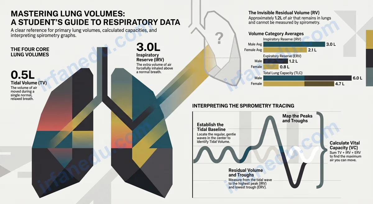
A lung volume chart displays the four primary lung volumes — tidal volume, inspiratory reserve volume, expiratory reserve volume, and residual volume — and the four lung capacities derived from them. Together they describe how much air the lungs can hold, move, and keep in reserve. Understanding these values is essential for biology, anatomy, health science, and any math course that includes data interpretation.
- 🫁 There are exactly 4 lung volumes and 4 lung capacities — and each capacity is just a sum of specific volumes.
- 📐 You can calculate any capacity if you know the right volumes — it’s straightforward addition.
- ⚠️ Residual volume cannot be measured by a basic spirometer — this is the most-tested exam fact.
- 📊 Average total lung capacity is ~6.0 L (male) and ~4.7 L (female).
⚡ TL;DR – Quick Summary
- 📌 4 lung volumes: TV (0.5 L), IRV (3.0 L), ERV (1.2 L), RV (1.2 L)
- 📌 4 capacities: TLC, VC, IC, FRC — each is a sum of 2–4 volumes
- 📌 Total Lung Capacity = TV + IRV + ERV + RV ≈ 6.0 L (male average)
- 📌 Vital Capacity = TV + IRV + ERV ≈ 4.8 L — the max you can breathe out
- 📌 Residual Volume cannot be measured by a standard spirometer
- 📌 Exercise increases TV dramatically; TLC stays the same
📑 Table of Contents
- Rule 1 — Know the 4 Lung Volumes Cold
- Rule 2 — Read the Master Lung Volume Chart
- Rule 3 — Understand the 4 Lung Capacities
- Rule 4 — Read a Spirometry Tracing Step by Step
- Rule 5 — Do the Math (Worked Examples)
- Rule 6 — Know How Age, Sex & Fitness Change the Chart
- Rule 7 — Spot the Common Mistakes
- 💡 Unique Insight
- 🧠 Quick Quiz
- 📝 Practice Problems
- ❓ FAQ
- 📚 Sources & References
Know the 4 Lung Volumes Cold
The four lung volumes are the foundation of every lung volume chart. Each one is a distinct, non-overlapping measurement — they do not share any air with each other.
| Volume | Abbreviation | Definition | Average (Adult Male) | Average (Adult Female) |
|---|---|---|---|---|
| Tidal Volume | TV (or VT) | Air moved in or out during one normal, relaxed breath | ~0.5 L (500 mL) | ~0.4 L (400 mL) |
| Inspiratory Reserve Volume | IRV | Extra air you can forcefully inhale above a normal breath | ~3.0 L | ~2.1 L |
| Expiratory Reserve Volume | ERV | Extra air you can forcefully exhale below a normal breath | ~1.2 L | ~0.8 L |
| Residual Volume | RV | Air remaining in lungs after a maximal forced exhalation — cannot be exhaled | ~1.2 L | ~1.1 L |
Read the Master Lung Volume Chart
The chart below is the single most important reference table for this topic. Bookmark it, print it, or copy it into your notes.
| Measurement | Type | Formula | Male Avg (L) | Female Avg (L) | Measurable by Spirometer? |
|---|---|---|---|---|---|
| Tidal Volume (TV) | Volume | — | 0.5 | 0.4 | ✅ Yes |
| Inspiratory Reserve Volume (IRV) | Volume | — | 3.0 | 2.1 | ✅ Yes |
| Expiratory Reserve Volume (ERV) | Volume | — | 1.2 | 0.8 | ✅ Yes |
| Residual Volume (RV) | Volume | — | 1.2 | 1.1 | ❌ No |
| Total Lung Capacity (TLC) | Capacity | TV + IRV + ERV + RV | 6.0 | 4.7 | ❌ No (needs RV) |
| Vital Capacity (VC) | Capacity | TV + IRV + ERV | 4.8 | 3.7 | ✅ Yes |
| Inspiratory Capacity (IC) | Capacity | TV + IRV | 3.5 | 2.5 | ✅ Yes |
| Functional Residual Capacity (FRC) | Capacity | ERV + RV | 2.4 | 1.9 | ❌ No (needs RV) |
Values are approximate averages for healthy adults. Individual values vary with height, age, sex, and fitness. Source: standard respiratory physiology references.
Understand the 4 Lung Capacities — They’re Just Sums
A lung capacity is always the sum of two or more adjacent lung volumes. Once you know that, you never need to memorize the formulas separately — you can rebuild them from logic.
| Capacity | Volumes It Includes | Plain-English Meaning |
|---|---|---|
| Total Lung Capacity (TLC) | TV + IRV + ERV + RV | The absolute maximum air the lungs can hold |
| Vital Capacity (VC) | TV + IRV + ERV | Max air you can move in one breath — your “usable” lung capacity |
| Inspiratory Capacity (IC) | TV + IRV | Max air you can inhale from a resting position |
| Functional Residual Capacity (FRC) | ERV + RV | Air left in lungs after a normal (not forced) exhalation |
In my experience, students who struggle with lung capacity questions are almost always trying to memorize formulas in isolation. The moment I show them that each capacity is just a stack of adjacent volumes on the spirometry graph — like stacking Lego bricks — everything clicks. Draw the stacked bar diagram once by hand and you’ll never forget it.
How Do You Read a Spirometry Tracing Step by Step?
Reading a spirometry graph is a skill, not just a lookup task. Follow these steps every time you see one on an exam.
- Find the resting baseline. The gentle, regular wave in the middle of the tracing is tidal breathing. The height of each wave = TV (~0.5 L).
- Identify the maximal inhalation peak. The highest point on the graph is the top of IRV. Measure from the top of the tidal wave to this peak = IRV.
- Identify the maximal exhalation trough. The lowest point the tracing reaches = bottom of ERV. Measure from the bottom of the tidal wave to this trough = ERV.
- Note the RV gap. The graph stops at the ERV floor. The air below that line (RV) is never shown on a basic spirogram — it’s invisible because it can’t be exhaled.
- Calculate capacities. Use the formulas: VC = TV + IRV + ERV; TLC = VC + RV; IC = TV + IRV; FRC = ERV + RV.
📈 Visual: Simplified Spirometry Tracing (ASCII)
Volume
(Litres)
  6.0 ┤                                        ← TLC (top)
      │   ╭──╮                                 ← IRV peak
  5.5 ┤   │  │
  5.0 ┤   │  │
  4.5 ┤   │  │
      │   ╰──╯ ← max inhalation (TV + IRV)
  4.0 ┤ ╭──╮    ╭──╮    ╭──╮   ← Tidal Breathing (TV ~0.5L each)
  3.5 ┤ │  │    │  │    │  │
  3.0 ┤ ╰──╯    ╰──╯    ╰──╯
      │
  2.5 ┤         ╰──────────╯   ← ERV (forced exhalation)
  1.5 ┤
      │ ▓▓▓▓▓▓▓▓▓▓▓▓▓▓▓▓▓▓▓▓  ← RV (~1.2L) — NEVER exhaled
  0.0 ┼──────────────────────────────────────
      │◄── TV ──►◄─── IRV ───►◄── ERV ──►│RV│
      │          Vital Capacity (VC)       │  │
      │          Total Lung Capacity (TLC)    │
Do the Math — 3 Worked Lung Volume Examples
Lung volume questions on exams are almost always straightforward arithmetic — but only if you know which formula to use. Here are three worked examples that cover the most common question types.
Given: TV = 0.5 L, IRV = 3.1 L, ERV = 1.1 L
Find: Vital Capacity (VC)
Formula: VC = TV + IRV + ERV
Calculation: VC = 0.5 + 3.1 + 1.1 = 4.7 L
Interpretation: This person can move a maximum of 4.7 L of air in one breath — slightly below the average male value of 4.8 L, which is normal variation.
Given: VC = 4.7 L (from Example 1), RV = 1.3 L
Find: Total Lung Capacity (TLC)
Formula: TLC = VC + RV
Calculation: TLC = 4.7 + 1.3 = 6.0 L
Interpretation: Total lung capacity is exactly at the average male value. The slightly higher RV (1.3 vs 1.2) is offset by the slightly lower VC.
Given: TLC = 5.9 L, TV = 0.5 L, IRV = 2.9 L, ERV = 1.1 L
Find: Residual Volume (RV)
Formula: RV = TLC − (TV + IRV + ERV)
Calculation: RV = 5.9 − (0.5 + 2.9 + 1.1) = 5.9 − 4.5 = 1.4 L
Interpretation: This is a slightly elevated RV. In a real clinical context, elevated RV can indicate air trapping (e.g., in asthma or emphysema).
How Do Age, Sex, and Fitness Change the Lung Volume Chart?
Lung volume values are not fixed — they shift with biological and lifestyle factors. Understanding these changes helps you interpret a lung volume chart correctly in real-world and exam scenarios.
| Factor | Effect on Lung Volumes | Which Values Change Most |
|---|---|---|
| 🧑 Sex (Male vs Female) | Males have larger lung volumes on average due to greater thoracic cage size | TLC ~6.0 L (M) vs ~4.7 L (F); VC ~4.8 L (M) vs ~3.7 L (F) |
| 📏 Height | Taller individuals have larger lungs; TLC increases with height | TLC, VC, IRV all increase |
| 🎂 Age | Lung volumes peak in early adulthood (~25 yrs) then slowly decline; RV increases with age | VC decreases; RV increases; TLC stays roughly stable until older age |
| 🏃 Exercise (acute) | TV increases dramatically during exercise; IRV and ERV decrease as reserves are used | TV can rise from 0.5 L to ~3.0 L; TLC unchanged |
| 🏋️ Athletic Training | Endurance athletes develop larger VC and stronger respiratory muscles | VC and IRV increase; RV may decrease slightly |
| 🚬 Smoking / COPD | Air trapping increases RV; VC decreases; FRC increases | RV↑, FRC↑, VC↓ — classic obstructive pattern |
| 🫁 Restrictive Disease | All volumes decrease proportionally (e.g., pulmonary fibrosis) | TLC↓, VC↓, RV↓ — all reduced |
The age-RV relationship is something I rarely see explained well in textbooks. As people age, their lung tissue loses elasticity — like an old rubber band. The lungs can’t recoil as forcefully, so more air gets trapped after exhalation. This is why RV creeps up with age even in healthy, non-smoking adults. It’s a normal physiological change, not a disease sign — and it’s a detail that separates a good exam answer from a great one.
What Are the Most Common Mistakes Students Make with Lung Volume Charts?
These are the errors I see most often — and they’re all avoidable once you know what to watch for.
| ❌ Common Mistake | ✅ Correct Understanding |
|---|---|
| Thinking RV can be measured by a spirometer | RV requires gas dilution or body plethysmography — it cannot be exhaled, so a spirometer cannot detect it |
| Confusing Vital Capacity with Total Lung Capacity | VC = TLC − RV. VC is the air you can move; TLC includes the air you can never move (RV) |
| Thinking TV stays the same during exercise | TV increases dramatically during exercise — it can rise 6x above the resting value |
| Mixing up IRV and ERV direction | IRV is above the tidal volume baseline (inhalation reserve); ERV is below it (exhalation reserve) |
| Assuming all capacities can be measured directly | Only capacities that exclude RV (VC, IC) can be measured by spirometry. TLC and FRC require RV measurement |
| Using male averages for female patients/data | Female lung volumes are consistently 20–25% smaller than male values — always check the sex of the subject |
💡 Unique Insight — What Most Lung Volume Guides Get Wrong
Almost every student resource presents lung volumes as static numbers to memorize. But here’s what most guides miss: the ratio of RV to TLC is clinically more important than the absolute values.
In a healthy adult, RV/TLC ≈ 20–25%. In a patient with emphysema, this ratio can climb above 40% because air trapping inflates RV while VC collapses. This ratio — not the raw RV number — is what physicians actually use to diagnose obstructive disease severity.
For students: if an exam question gives you both RV and TLC, always calculate RV/TLC × 100. A ratio above 35% in a question scenario almost always signals a pathological condition. No other student guide at this level teaches this ratio as a decision tool.
🧠 Quick Quiz — Test Your Lung Volume Knowledge
1. A person has TV = 0.5 L, IRV = 3.0 L, ERV = 1.2 L, RV = 1.3 L. What is their Total Lung Capacity?
2. Which lung volume CANNOT be measured by a standard spirometer?
Sources & References
Written and fact-checked by Dr Irfan Mansuri. External links open in a new tab and are provided for further reading and verification.